
Cutting Off The Mutation
In 2014, during a networking break in an event, I commented with a researcher: “I´m just waiting for someone to find a way to cut off the mutation in the DNA and correct it.”. And finally, this idea as a chance to come true!
The new future is here: Clustered regularly-interspaced short palindromic repeats (abbreviated as CRISPR, pronounced as crisper)
CRISPR-Cas9 is a genome-editing technique, like a genetic “cut and paste”, that can be roughly compared to a film editing machine. When a film has a wrong shot, it can be cut in a precise location and it can be replaced with another shot.
But before explaining what CRISPR-Cas9 does, some highlights about the genetic code are necessary.
The Genetic Code
It is the set of rules by which information encoded within the genetic material (DNA or RNA sequences) is translated into proteins by living cells. Proteins are macromolecules, which means that they are made with sequences of smaller molecules, in this case amino acids, of which human cells use 20. So proteins are, in essence, sequences of these 20 different molecules of the same class that interact with each other to form complex structures, the proteins themselves. Proteins can also interact with each other and other molecules to form even more complex structures, making up most of our cellular structures [1].The genome (the set of all the DNA of an organism, which includes all genes) exists in each cell, inside the nucleus, in the form of nucleotides (a block of one nitrogenous base, a five-carbon sugar and one phosphate group), which are the building blocks of the nucleic acids: DNA and RNA. Most DNA molecules consist of two strands of nucleotides coiled around each other to form a double helix. In DNA the two strands of nucleotides form base pairs (unit: bp), by binding to each other [2].
There are 4 different nucleobases (nitrogenous base of the nucleotide) in DNA: Adenine, Guanine, Cytosine, Thymine. In RNA Thymine is substituted by Uracil. The base pairs always form with the same bonds: Adenine always binds to Thymine (Uracil in RNA) and Guanine always binds to Cytosine [2].

| Credits: Science Prof Online |
The genetic code is defined by sequences of groups of three nucleotides, called codons, that specify which amino acid will be added to the sequence during protein synthesis, for which a form of RNA is an intermediate (messenger RNA carries the gene's information outside of the nucleus for protein production). With some exceptions, a three-nucleotide codon in a nucleic acid sequence specifies a single amino acid, but some amino acids can be specified by more than one codon (making it possible to define a specific amino acid sequence from the DNA sequence, but not the exact DNA sequence from a protein).
The total length of the human genome is over 3 billion base pairs [3]. It is organized into chromosomes, complexes of DNA coiled around proteins called histones [4]. Humans have 22 paired chromosomes (autosomes), plus two X chromosomes in females or a Y and an X chromosome in males (the allosomes, also known as sex chromosomes) [3].

|
There are many types of RNA. It acts as a regulator or temporary storage molecule, but does not store genetic infromation except for taking it out of the nucleus in the form o messenger RNA (mRNA), and in certain viruses.
Credits: Hyperphysics.edu |
The mutation that causes achondroplasia is a single point mutation, a transition guanine-to-adenine (1138G-A) at nucleotide 1138 of the FGFR3 gene, located in chromosome 4 (nucleotide 1138, which is normally a guanine, is substituted by an adenine). This transition produces a nucleotide change which, in turn substitutes the amino acid glycine for the amino acid arginine in the 380th position in the protein(the substituted glycine was the 380th amino acid and is written as Gly380Arg or G380R) [5]. This means that the mutation in achondroplasia is due to a change in a single “letter” (nucleobase) in that single gene.
|
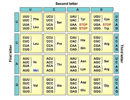
The genetic code. This image shows which codons code for which amino acids and includes the codons the codons that signal for one of the phases of protein synthesis to stop, the translation.
Credits: nptel.ac.in |
CRISPR-Cas9
CRISPR-Cas9 is a technique invented by Dr. Jennifer Doudna, Dr. Emmanuelle Charpentier and Dr. Feng Zhang (there is a lot of confusion about who invented it first and who holds the patent of this technology). It is inspired by the defensive system of some bacteria against attack by other pathogens (other bacteria, virus) [6, 7].
These enzymes detect a difference between the genetic material of bacteria and viruses and destroy the latter (viruses have a specific signal in their genetic material). The technique has transformed the natural machinery of these bacteria into a programmable tool that can cut any segment of DNA and replace a mutated piece that normally causes a disease. Like a word processor using “cut and paste”, this tool finds the desired piece of DNA and replaces it with its corrected version, using a molecular guide (made of RNA) [8, 9].
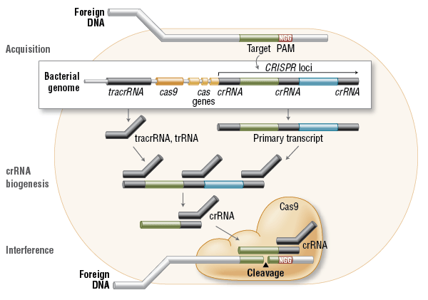
| In the acquisition phase, foreign DNA is incorporated into the bacterial genome at the CRISPR loci (a locus is where a gene is situated in a chromosome). CRISPR loci are then transcribed and processed into crRNA during crRNA biogenesis. During interference, Cas9 endonuclease complexed with a crRNA and separate tracrRNA cleaves foreign DNA containing a 20-nucleotide crRNA complementary sequence adjacent to the PAM sequence (the specific viral signal). (Figure not drawn to scale.) Credits: New England BioLabs. |
“The genome-editing method called CRISPR has matured into a molecular marvel that much of the world—not just biologists—has noticed, which is why it has been selected Science’s 2015 Breakthrough of the Year, revealing its true power in a series of spectacular achievements. Two striking examples—the creation of a long-sought “gene drive” that could eliminate pests or the diseases they carry, and the first deliberate editing of the DNA of human embryos—debuted to headlines and concern. The embryo work (done in China with nonviable embryos from a fertility clinic) even prompted an international summit this month to discuss human gene editing. The summit confronted a fraught—and newly plausible— prospect: altering human sperm, eggs, or early embryos to correct disease genes or offer “enhancements.”

| Image credits: The Wall Street Journal. |
A paper with high relevance for achondroplasia was published by Wojtal el at.:
Versatility of CRISPR/Cas9 for Developing Treatments for Inherited Disorders, (Wojtal et al., Spell Checking Nature: The American Journal of Human Genetics (2016), http://dx.doi.org/10.1016/j.ajhg.2015.11.012), in which can be read the following:
“Here, to investigate the therapeutic potential of CRISPR/Cas9 in a diverse set of genetic disorders, we establish a pipeline that uses readily obtainable cells from affected individuals. We demonstrate preferential elimination of the dominant-negative FGFR3 c.1138G>A allele in fibroblasts of an individual affected by achondroplasia.”
“We then electroporated the achondroplasia fibroblasts with Cas9 and FGFR3 sgRNA 1 and used deep sequencing to analyze the FGFR3 exon 9 region...The data demonstrate that our designed sgRNA preferentially targets the mutant allele“
“The CRISPR/Cas9 system provides a rare opportunity to employ a technology that can not only target the underlying primary disease-causing genetic abnormalities but also alter genetic modifiers that play a critical role in the pathogenesis of a certain disease. Here, we have developed a pipeline in which genome-engineering strategies use easily accessible cells from affected individuals and provide evidence of the versatility of the CRISPR/Cas9 system for various genetic conditions.”
The first steps have been taken! I´m sure that from now on, more studies using CRISPR/Cas9 will be published and that a new therapy for achondroplasia will rise.
Sources
1. Berg JM, Tymoczko JL, Stryer L. Biochemistry. 5th edition. New York: W H Freeman; 2002. Section 3.1, Proteins Are Built from a Repertoire of 20 Amino Acids. Available from: https://www.ncbi.nlm.nih.gov/books/NBK22379/ 2. Berg JM, Tymoczko JL, Stryer L. Biochemistry. 5th edition. New York: W H Freeman; 2002. Section 1.1, DNA Illustrates the Relation between Form and Function. Available from: https://www.ncbi.nlm.nih.gov/books/NBK22415/
3. Brown TA. Genomes. 2nd edition. Oxford: Wiley-Liss; 2002. Chapter 1, The Human Genome. Available from: https://www.ncbi.nlm.nih.gov/books/NBK21134/.
4. National Library of Medicine. What is a chromosome? Cells and DNA 2017 [cited 2017 20/06]; Available from: https://ghr.nlm.nih.gov/primer/basics/chromosome.
5. McKusick, V.A.H., Patricia A. . 134934 FIBROBLAST GROWTH FACTOR RECEPTOR 3; FGFR3. 2015 [cited 2017 20/06]; Available from: https://www.omim.org/entry/134934#creationDate.
6. Kupecz, A. (2014). Who owns CRISPR-Cas9 in Europe? Nature Biotechnology, 32(12), 1194–6. Available from: http://search.proquest.com/openview/c3c08e385519821edc4d14fdc649daee/1?pq-origsite=gscholar&cbl=47191
7. Ledford, H. (2017). Five big mysteries about CRISPR’s origins. Nature, 541(7637), 280–282. Available from: http://doi.org/10.1038/541280a
8. Song, G., Jia, M., Chen, K., Kong, X., Khattak, B., Xie, C., ... Mao, L. (2016). CRISPR/Cas9: A powerful tool for crop genome editing. The Crop Journal, 4(2), 75–82. http://doi.org/10.1016/j.cj.2015.12.002
9. Cyranoski, D. (2016, November). CRISPR gene-editing tested in a person for the first time. Nature. England. http://doi.org/10.1038/nature.2016.20988
10. Travis, J. (2015). Making the cut. Science, 350(6267), 1456 LP-1457. Retrieved from http://science.sciencemag.org/content/350/6267/1456.abstract
11. Wojtal, D., Kemaladewi, D. U., Malam, Z., Abdullah, S., Wong, T. W. Y., Hyatt, E., ... Cohn, R. D. (2016). Spell Checking Nature: Versatility of CRISPR/Cas9 for Developing Treatments for Inherited Disorders. American Journal of Human Genetics, 98(1), 90–101. http://doi.org/10.1016/j.ajhg.2015.11.012

