This web site uses cookies. Do you accept the use of external cookies? Yes No Know more x
Survey about achondroplasia
We have designed a questionnaire with the purpose to better know the population connected to achondroplasia.
The questionnaire is optional and anonymous. The data obtained will be used for statistical purposes and to better understand the natural history of achondroplasia. Please consider answering.
If you would like to take the survey later, you can click on the link at the top "Register at BA"

Adipocytes, also known as fat cells, are the cells that primarily compose adipose tissue, specialized in storing energy as fat.

Yellow_adipose_tissue_in_paraffin_section_-_lipids_washed_out
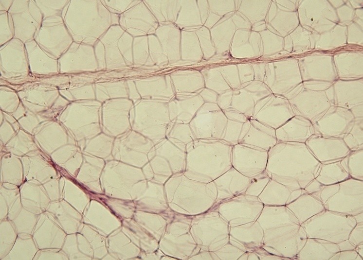
"Yellow adipose tissue in paraffin section - lipids washed out". Licensed under CC BY-SA 3.0 via Wikimedia Commons


Bone marrow is the flexible tissue in the interior of long  bones.  The two types of bone marrow are "red marrow", which consists mainly of hematopoietic tissue, that originates blood cells: erythroid cells (red blood cells) , lymphocytes (white blood cells) and myelocytes (as platelets), and "yellow marrow", which is mainly made up of fat cells.

"619 Red and Yellow Bone Marrow" by OpenStax College - Anatomy & Physiology, Connexions Web site. http://cnx.org/content/col11496/1.6/, Jun 19, 2013.. Licensed under CC BY 3.0 via Wikimedia Commons - http://commons.wikimedia.org/wiki/File:619_Red_and_Yellow_Bone_Marrow.jpg#mediaviewer/File:619_Red_and_Yellow_Bone_Marrow.jpg
A femoral head with a cortex of cortical bone and medulla of trabecular bone. Both red bone marrow and a central focus of yellow bone marrow are visible. "619 Red and Yellow Bone Marrow" by OpenStax College - Anatomy & Physiology, Connexions Web site

"Fat embolism syndrome (FES) was first described by Von Bergmann in 1873 in a patient with a fractured femur. While fat within the circulation (fat embolism) is relatively common following long-bone fracture, the clinical pattern of symptoms that make up FES is less so, occurring in 1% to 3% of isolated long-bone fractures and 5% to 10% of patients with multiple skeletal trauma
." (Excerpt from the American Journal of Orthopedics)

"Fat embolism syndrome (FES) is rare in pediatric patients; it is approximately 100 times less likely to develop in children and adolescents than in adults. The mortality rate for FES in children ranges from 1% to 15%, somewhat lower than that in adults (10%-20%). FES most often occurs after fractures of the long-bones, primarily the femur and pelvis. The development of FES after intramedullary nailing of long-bones in adults is well-described, with identified risk  factors of advanced age, polytrauma, prolonged immobilization, and male sex. To our knowledge, there have been no documented cases of FES after isolated closed tibial shaft fractures but before operative treatment." (Excerpt from the case report "Fat Embolism Syndrome in an Adolescent Before Surgical Treatment of an Isolated Closed Tibial Shaft Fracture" R. Sawyer, et al, Dec 2012).


And how can this happen in a lengthening surgery?

Most likely, fat cells/globules are dislodged/released from the bone marrow while drilling the bone during surgery and enter a small vessel of the bone (vascular sinusoids) that will lead to the lungs, brain and heart, producing obstruction.

What are the signs?

This process can take 1 to 3 days to develop with progressive respiratory changes, neurological symptoms and skin rash and can end up in death.

In a very conclusive article:


"Several methods are available for progressive limb lengthening, including centromedullary nailing, external fixation, or a combination. Each technique has its own advantages and drawbacks. In trauma victims, use of centromedullary nailing is associated with potentially fatal fat embolism. This fatal outcome might also occur during limb lengthening, particularly in bilateral procedures. To our knowledge, fat embolism has not been reported with the use of centromedullary nail for limb lengthening...   Fat embolism could result from several factors, as reported in the literature. While the bilateral nature of the procedure has been incriminated, the observation of an embolism during a unilateral procedure suggests other factors may be involved. Considerable increase in endomedullary pressure during reaming and insertion of the nail has been demonstrated. At the same time, there is the question as to whether the reduction of the diminution of medullary pressure by corticotomy would be an efficient way of reducing the risk of fat embolism. Based on the analysis of our three cases, we suggest that the best way to avoid fat embolism might be to drill several holes within the area of the osteotomy before reaming, in order to reduce endomedullary pressure. This can be achieved via a short skin incision, sparing the periosteum before low energy osteotomy. Since applying this protocol, the three centers have implanted 17 lengthening nails, without a single case of fat embolism."

Fat embolism during limb lengthening with a centromedullary nail: three cases,  Bollini G et al,  Rev Chir Orthop Reparatrice Appar Mot. 2008 Sep:

Lastly, the risk of fat embolism is real in limb lengthening, but this surgery exists since 1954 (developed by Dr.  Gavriil Ilizarov) and nowadays is executed in a much safer way and with more advanced techniques.
To view this site you need Internet Explorer 8 (or a higher version)
or another internet browser.


Thank You.