Osteopathy proposal for prevention of otitis media in achondroplasia
Mauro Banfi is a physiotherapist who also has a degree in osteopathy, from the College Osteopathique de Provence - Marseille, France.
I met him recently during the AISAC congress. AISAC is the Italian Association for the Information and Study of Achondroplasia. In this event, Mauro presented a proposal for the prevention of otitis media by using osteopathy techniques and later he did a practical workshop, explaining some of the techniques in a young child with achondroplasia. Here we share a video of Mauro Banfi applying osteopathy techniques to prevent otitis media in achondroplasia.
In children, the Eustachian tube is horizontal, shorter and "... in a child with achondroplasia, is also short and small. Because these children are quite hypotonic, they may spend the first year of life lying in bed, meaning more pronounced horizontal position, that leads to the marked increase of otitis media and the accumulation of fluid in the middle ear seen in childhood in achondroplasia". (Benedetto Nicoletti, Elio Ascani, Victor A. McKusick, Human Achondroplasia: A Multidisciplinary Approach, 2012)
The Eustachian tube consists of a bony part and a cartilaginous part. A portion (1/3) of the tube proximal to (same as next to) the middle ear is made of bone. Usually, the tube is collapsed (closed), but it opens when swallowing or with positive pressure.
- ventilation and pressure regulation of the middle ear
- protection of the middle ear from nasopharyngeal secretions and sound pressures
- clearance and drainage of middle ear secretions into the nasopharynx
Specific muscles (the levator veli palatini and the tensor veli palatini) cooperate to open the tube regularly with a traction on a cartilaginous structure (these muscles pull the structure made of cartilage). But in a child with achondroplasia, these muscles do not have the same efficacy: the Eustachian tube does not accomplish an active natural opening and this leads to ear infections and fluid build-up in the middle ear cavity. This occurs because the tube does not open appropriately, causing mainly inflammation and infection, pain, rise of pressure, tympanic membrane rupture (bulging eardrum) and fever.

| Image: Eurolab |

|
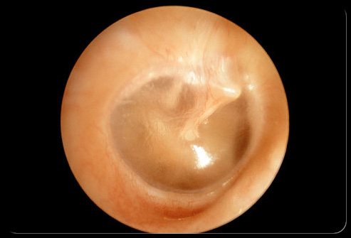
This is a tympanic membrane (eardrum) in tension, with fluid inside (the fluid is in the middle ear cavity). Image taken from the ear canal (outside to the inside). Credits: Eurolab. |
In many cases of chronic otitis media, a ENT doctor has to perform a myringotomy, which means that he places a grommet, in a precise location of the tympanic membrane (eardrum) to allow drainage of middle ear infections that occur.
Another way to reduce the frequency of otitis media in achondroplasia is shown in Muaro Banfi's video.
Point 1. The first idea is that Prevention is the key. So, action must be taken before there's signs of otitis. Next is the video where Mauro Banfi executes stretching techniques in order to open the eustachian tube, allowing natural drainage of the middle ear.
Point 2. Please, don't perform these techniques by yourself and without professional supervision. Mauro Banfi can eventually be contacted by your physiotherapist or osteopath for evaluation of clinical cases.
Point 3. These techniques are complementary to the ENT approach, so they don't substitute going to the doctor.
Point 4. Some sounds when pronounced have the ability to produce the opening of the tube. You can teach the child to produce these sounds, playing and singing together and this is easy and not invasive. K and GH. E and I.
A special Thank you to Mauro Banfi for editing this video.


